Your list is empty
Add products to request a quote

Imagine you have two identical keratinocytes kept together in an incubator in 0.03 mM calcium and 1.2 mM calcium. After a day or so, the cells in the second bottle flatten, stick to their neighbors, and activate genes that did not play a role in the first. Just a simple change in calcium lets you watch a skin barrier build itself on a plastic dish.
If you use keratinocytes, you probably asked questions about what markers to follow and when they show up. You probably also wanted to know how long until you harvest your cells, or if you should use primary cells that resemble donor skin, or HaCaT cells that will not die.
This guide answers each question, adds a protocol to switch calcium, a comparison of models, a checklist of reagents, and standard detection tips.
Keratinocyte differentiation occurs when a basal cell that has been rapidly dividing ceases to divide, grows up, and produces genes for keratins, involucrin, loricrin, and filaggrin. Cells change shape, produce different proteins, and develop features of the mature epidermis.
As a result, there’s barrier formation, less water loss, and underlying tissues are protected. Understanding what are keratinocytes and how they work explains why they are so important for skin health.
Researchers also study keratinocyte differentiation in laboratory experiments to understand the development of the skin, disease, and reaction to experimental treatments.
The Stages: basal → spinous → granular → cornified
Cells pass through four layers. Basal cells divide, spinous cells build strong attachments, granular cells assemble the protective envelope, and cornified cells form the barrier.
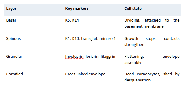
Basal keratinocytes divide to maintain the epidermis. As daughter cells ascend, they slow down proliferation and express proteins associated with further differentiation.
In the granular layer, cells secrete proteins and lipids that contribute to barrier formation; they undergo terminal differentiation, producing the cornified layer of flattened, nonviable corneocytes.
The life cycle of a keratinocyte is a coordinated process of proliferation, maturation, and shedding. Many aspects of this process are recapitulated in in vitro cultures, but a monolayer can't fully recapitulate the details of intact skin.
Keratinocyte differentiation markers can be used to assess cell state by gene expression, protein abundance, and cell distribution. One marker alone is insufficient.
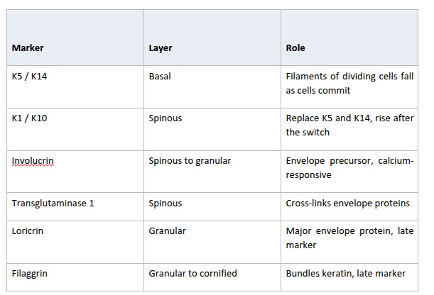
The expression of markers may depend on the model, culture conditions, and sampling time; for example, K1 and K10 expression may increase before late markers such as loricrin and filaggrin become prominent.
Researchers should also consider the distinction between gene expression and protein abundance. A rise in messenger RNA does not always produce an equivalent increase in protein. Combining molecular and imaging methods can help establish whether the cells are progressing through the expected stages.
The calcium switch of keratinocytes is when cells are switched from a low calcium condition to a medium with more extracellular calcium. The cells attach themselves to one another and are signaled to differentiate.
Calcium regulates the formation of junctions between nearby keratinocytes as well as the signaling processes involved in maturation. Researchers often rely on controlled calcium changes to study these responses under laboratory conditions.
Choose primary cells when donor relevance and full terminal differentiation matter. Choose HaCaT cells when you need an unlimited, reproducible supply.
Choose primary cells when your research depends on normal human keratinocyte behavior, donor-specific responses, or physiological relevance. Primary cells have limited growth potential, exhibit donor-to-donor variation, and may be sensitive to culture conditions.
HaCaT cells are useful when you need a human keratinocyte model for repeated experiments. They have been widely used in skin biology, wound healing, and differentiation studies. Because HaCaT cells are immortalized, they do not need to be treated the same way as primary cells.
XL Biotec supplies HaCaT cells in Thailand as a frozen cryovial of an adherent human keratinocyte cell line, ready to thaw and use in the routine above. Order yours today and start your consistent cultures.
Culture conditions are crucial for maintaining keratinocyte health, cell growth and differentiation. Before performing your experiment, make sure that your culture media, serum, supplements, and calcium levels are adequate for your chosen cell model. Use the following checklist to prepare your cell culture workflow:
Depending on the lab, HaCaT culture conditions may vary. Ensure you follow the supplier’s recommendations and establish the right conditions before starting the differentiation study.
Choosing suitable culture vessels, sterile consumables, and supporting reagents is equally important for establishing a consistent workflow.
XL Biotec supplies HaCaT cells in Thailand and a range of tissue culture products that researchers can explore when sourcing supplies for their laboratory needs.

Use qPCR for early transcript changes, Western blot for protein levels, and immunofluorescence to see where proteins sit.
With qPCR, measure KRT1, KRT10, IVL, and TGM1 early and LOR and FLG late. For Western blot, probe K5 or K14 next to K1 or K10 to catch the keratin swap. For immunofluorescence, stain involucrin and loricrin, and include a DAPI counterstain to confirm that cells are stratifying.
Consistent and accurate results start with the right cell model, well-controlled culture conditions, and appropriate markers to address your research question. If your study relies on normal responses in human cells, primary keratinocytes will provide an ideal model. And if you require reproducible cells, HaCaT is your choice.
XL Biotec supplies HaCaT cells in Thailand, alongside other tissue culture supplies. If you are planning a differentiation experiment, request a HaCaT quote or contact the XL Biotec team to discuss product specifics, culture requirements, and the reagents needed for your research.
Keratinocyte differentiation occurs when basal cells stop dividing and gradually move toward the skin's surface. During this process, they produce proteins such as K1, K10, involucrin, loricrin, and filaggrin before becoming flat corneocytes.
K5 and K14 are commonly used to identify basal keratinocytes, while K1 and K10 are used to identify spinous cells. Involucrin, loricrin, and filaggrin are associated with the later stages of differentiation, particularly the granular and cornified layers.
A calcium switch involves increasing calcium levels in the culture medium to encourage differentiation. This may begin at or above approximately 0.1 mM, although many laboratory protocols use approximately 1.2 mM calcium.
Early differentiation markers such as K1, K10, and involucrin increase within 24–48 hours. Cells are collected after 3–5 days, depending on the experiment setup.
Primary keratinocytes are isolated from donors and have a limited lifespan in culture. HaCaT cells are immortalized, allowing them to be maintained and expanded over many passages.
HaCaT cells can differentiate after a calcium switch and express more K1, K10, and involucrin. But loricrin and filaggrin levels may remain low in some culture conditions.
Serum-free medium is the standard for keratinocyte culture. HaCaT cells are usually cultured in high-glucose DMEM with 10% fetal bovine serum.
qPCR tests for changes in gene expression, Western blot tests for changes in protein expression, and immunofluorescence reveals where in the cell the protein is located. Together, they give a more complete picture of differentiation.
Differentiation refers to the entire process of final transformation of basal keratinocytes into corneocytes. Keratinization is the second part of this process, in which keratin is deposited and the protective skin barrier forms.
XL Biotec supplies HaCaT cells in Thailand, as well as DMEM, fetal bovine serum, and tissue culture reagents. Contact the company at info@xlbiotec.com for further information.
