Your list is empty
Add products to request a quote

The idea of growing living cells outside the body is like something out of a science fiction movie. What once seemed futuristic has now become an everyday reality in laboratories around the world, as tissue culture has become one of the most routine and essential techniques in modern biological research.
Whether you are a newcomer to the field of cell biology or you are looking to refine your existing understanding of the field, understanding the techniques behind successful tissue culture will ensure that you are achieving the most accurate results from every experiment you perform.
Here is the full picture of tissue culture, from the fundamental role of enzymes like trypsin to the specialized requirements of stem cell culture.
Growing cells outside the body in a controlled laboratory environment is precisely what tissue culture entails, ranging from individual cells to whole tissues and organs. Scientists can sustain these cells in carefully designed containers, such as flasks, plates, or multi-well plates, which provide them with everything they need to thrive in the body.
The application of tissue culture ranges from almost all areas of life science research. Pharmaceutical firms use it to test proposed drugs on human cell cultures before testing on animals. Vaccine makers use it to grow viruses in cell cultures to produce enough for immunization campaigns.
Cancer researchers study how cancer cells act and why they are susceptible to drugs. Developmental biologists study how stem cells develop into specialized tissues. In all these areas, the quality of the method and the reagents used is what makes the science.
A proper understanding of tissue culture requires not only an understanding of the larger picture but also the details that comprise it, such as the media that nourish the cells or the enzymes that release them from the growth surface.
Cells in the body receive a constant supply of nutrients, hormones, and growth factors through the bloodstream. In the laboratory, tissue culture media supply these nutrients. These nutrient solutions provide a precisely defined combination of amino acids, vitamins, salts, glucose, and buffering agents.
Selecting the appropriate medium for your cells is one of the most critical decisions you make in any tissue culture experiment. Different cell types have distinct metabolic requirements, and the use of inappropriate media can result in reduced growth rates, altered cell behavior, or cell death. This explains why there are so many different formulations available in the market, each designed for a specific purpose.
Among the various cell culture media, DMEM/F12 medium is the most versatile for cell biology research. DMEM/F12 is a hybrid medium that consists of Dulbecco’s Modified Eagle’s Medium and Ham’s F-12 Nutrient Mixture. The medium has been found helpful for supporting various cell types, including epithelial cells, fibroblasts, and, notably, stem cells. Also, the medium’s nutrient content is balanced to support serum-free cell culture.
On the other hand, RPMI 1640 media plays an equally important role in cell biology research. Researchers originally developed this formulation to culture human leukemic cells, and it has since proven especially effective for suspension cell cultures, particularly lymphocytes and other immune cell types. The medium has a higher concentration of bicarbonate and other amino acids to meet the metabolic requirements of floating cells suspended in the medium rather than attached to the cell culture flask surface.
Beyond the growth medium itself, successful tissue culture requires a set of ancillary cell culture reagents that handle everything from maintaining sterility to preparing cells for analysis. Knowing what each reagent does and why it’s important will help you create a more efficient workflow.
Phosphate-buffered saline, or PBS, is perhaps the simplest yet most commonly used reagent in any cell culture lab. Scientists use it to wash cells before passaging, to remove serum before adding trypsin, and to resuspend cells for counting or analysis. Its isotonic formulation prevents osmotic stress on cells during these handling steps.
Antibiotics such as penicillin and streptomycin serve to protect cultures from bacterial contamination, which can quickly ruin an experiment in a matter of hours if left unchecked. Antifungal agents such as amphotericin B can also be used to protect cultures from fungal contamination.
While proper sterile technique is still the best first line of defense, these reagents are an important last resort, especially in busy labs where multiple individuals share incubators and biosafety cabinets.
L-glutamine is another crucial supplement because it is a major energy source for many rapidly dividing cell types. It degrades over time in solution, so investigators who make their own complete media must add it fresh or use the more stable dipeptide form, GlutaMAX, to achieve consistent performance in their experiments.
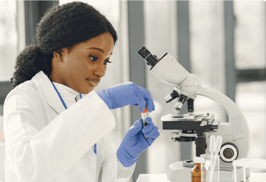
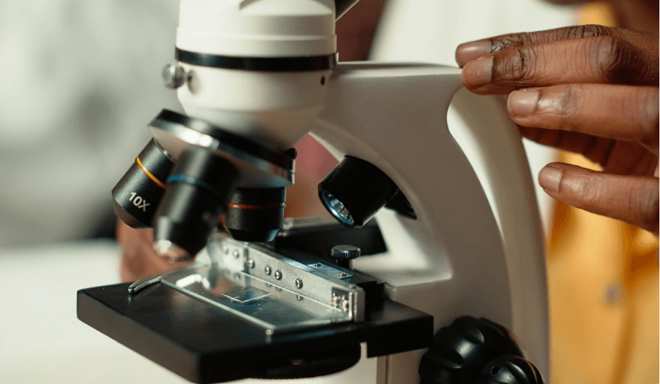
Cell passaging ranks among the most routine tasks in tissue culture, in which cells are transferred from one vessel to another as they approach confluency. Most cells are adherent, meaning that they stick well to the tissue culture surface through protein-based bonds, and you need a way to release them without damaging them.
This is where trypsin powder enzyme comes into play, becoming an indispensable tool in tissue culture work. Trypsin is a serine protease that cleaves peptide bonds on the carboxyl side of lysine and arginine residues.
This means it breaks down proteins that hold cells in place, releasing them into suspension so they can be counted and reseeded into a fresh tissue culture vessel. When tissue culture scientists make trypsin solutions from trypsin powder, they usually prepare a PBS solution at 0.25 percent, sometimes with EDTA to chelate calcium ions involved in cell adhesion.
The trick to effectively carrying out trypsinization is getting the timing right. Over-trypsinization of cells may damage cell surface receptors or membrane proteins, affecting cell behavior or viability. On the other hand, under-trypsinization of cells makes them difficult to count or reseed because they stick together. Most adherent cell lines take between two and five minutes at 37°C to detach from the cell culture flask or surface.
Different research questions call for different cell models, and understanding the characteristics of the most commonly used cell lines for culture will help you choose the right system for your research needs.
WPMY-1 cells, for example, demonstrate the value of specialized cell lines tailored to different research questions. Researchers use these immortalized human prostatic stromal myofibroblast cells extensively to study prostate biology, stromal-epithelial interactions, and the tumor microenvironment in prostate cancer.
Because the cells retain the characteristics of the original tissue, they are a better alternative to other fibroblast cultures for research on prostate disease and the development of therapeutic strategies.
Other commonly used cell lines include HeLa cells, the first and most widely used human cell line, derived from cervical cancer tissue and used for a wide variety of molecular biology and cell biology applications.
The HEK293 cell line, derived from human embryonic kidney tissue, has also become a workhorse for protein expression and transfection. Vero cells, derived from African green monkey kidney tissue, remain the most commonly used cells in virology and vaccine production.
No tissue culture lab functions without a plan for long-term cell storage. Experiments end, projects pause, and researchers must bank cell lines safely so work can resume without starting from scratch. Cryopreservation addresses this issue by cooling cells to very low temperatures where biological processes are effectively halted, allowing cells to be stored indefinitely until they are required.
The cryopreservation procedure must be performed carefully, with the cryopreservation medium protecting cells during freezing. Regular cell culture media cannot protect cells from ice crystal formation during freezing, which physically disrupts cell membranes and cell integrity.
Cryoprotective agents, typically dimethyl sulfoxide (DMSO), enter cell membranes and inhibit ice crystal formation by lowering the freezing point of cellular fluids. A standard cryopreservation medium consists of 10% DMSO in the base medium or in serum.
Nevertheless, some sensitive cell types, particularly stem cells and primary cultures, benefit from specialized commercially formulated cryopreservation solutions that optimize recovery rates and post-thaw viability.
The rate at which the cells are frozen is equally important to the composition of the freezing medium. Cells frozen at too rapid a rate will form ice crystals. At the same time, those frozen at too slow a rate will experience osmotic problems because water will leave the cells faster than the cryoprotectant can enter to counteract the effect.
The highest likelihood of cells surviving the process intact will be achieved using a controlled-rate freezer or a container of isopropanol at a cooling rate of 1°C/min.
Stem cells are among the most intriguing and challenging aspects of tissue culture. Their ability to renew and differentiate into any cell in the human body makes them extremely important in regenerative medicine and drug discovery.
At the same time, they are among the most sensitive cells in culture and require very specific conditions that may not always be met in a standard tissue culture environment. The stem cell culture substrate plays an important role in determining whether they remain undifferentiated or begin to differentiate.
In the body, stem cells sit within a specialized microenvironment called the niche, which provides both biochemical signals and physical support through the extracellular matrix. This microenvironment supplies stem cells not only with biochemical signals but also with mechanical support through the extracellular matrix.
Therefore, to create an environment in the lab that supports stem cells, it is important to use a substrate that mimics the natural properties of the matrix. Matrigel has traditionally been the substrate of choice for human pluripotent stem cells, providing a complex mixture of extracellular matrix proteins, including laminin, collagen, and entactin.
Nevertheless, its animal origin and batch-to-batch inconsistency have fueled the need for more defined, xeno-free alternatives. Recombinant fragments of laminin and vitronectin, as well as synthetic polymer-based matrices, have recently provided scientists with more consistent and translationally friendly options for stem cell expansion.
In addition to the choice of substrate, stem cell culture also requires specific media compositions, which are usually serum-free and defined to meet growth factor requirements, promoting self-renewal without inducing differentiation.
DMEM/F12 is the foundation for most of these media compositions, which are further supplemented with defined growth factors, such as bFGF for pluripotent stem cells or particular cytokine mixtures for directed differentiation approaches.
The quality of your tissue culture techniques will always reflect the quality of the tissue culture supplies you use. Inconsistent media formulations, trypsin preparations, or cryopreservation solutions will always be problems that no amount of technique can overcome.
Getting tissue culture supplies from reputable distributors that have proper cold chain management practices will ensure that your research techniques are protected from the very beginning.
Researchers looking for a reliable source of tissue culture media, cell culture reagents, and other specialty products, such as stem cell culture substrates, will find the greatest benefit from tissue culture suppliers that offer both product depth and expertise.
Having a reliable source of tissue culture supplies to turn to when you have questions or concerns will help you conduct your research techniques more efficiently.
Tissue culture is at the center of all biological research, and every aspect of the system counts. The medium you choose, the enzyme you use to facilitate cell passage, the substrate you provide for stem cell expansion, and the cryopreservation method you select all affect whether your cultures succeed or fail.
Creating a sound knowledge base for each aspect and making informed decisions about your reagents will give your research a solid foundation, helping you produce results you can bank on. From the initial flask of adherent cells to the most intricate stem cell differentiation protocol.
DMEM/F12 combines two media formulations into one balanced hybrid that suits a wide range of adherent cell types and works particularly well for stem cell culture under serum-free conditions. RPMI 1640 was developed specifically for suspension cells, especially lymphocytes and other immune cell types, and its formulation reflects the distinct metabolic needs of these cells compared to those of adherent lines.
The right substrate depends on your cell type, your downstream application, and whether your work requires xeno-free conditions.
Poor detachment usually occurs because the trypsin solution has lost activity due to repeated freeze-thaw cycles or improper storage, because residual serum in the well inhibits the enzyme, or because the incubation time or temperature is insufficient.
