Your list is empty
Add products to request a quote

Key Takeaways:
Tissue culture contamination is a major threat to microbiology experiments.
When a nutrient medium is infected, the invaders can alter cell behavior and lead to inconclusive results. Some contaminants may destroy experiments completely and cause massive resource wastage.
Tissue culture contamination can also derail critical experiments, prolonging the duration required to develop important vaccines or drugs. Not the ideal scenario when researchers are racing against time to push back a raging pandemic or terminal illness.
Considering the critical role of tissue culture in modern biotechnology, even a single instance of contamination is one too many. Fortunately, you can implement certain measures to avoid these costly mishaps.
Read on for more insights into tissue culture contamination.
Table of Contents:
Tissue culture contamination happens when unwanted agents invade a nutrient culture medium, altering plant or mammalian cell growth. The invaders can be biological (such as bacteria) or chemical (such as detergent residues).
According to estimates, about 11% of all mammalian cell cultures get contaminated by invading agents like mycoplasma. Contamination can result in costly repercussions, including;
Human error accounts for the vast majority of cell culture contamination. Common routes include;
In fact, even seemingly benign actions like talking over cell cultures can contaminate a nutrient medium. This happens when you inadvertently transmit unwanted salivary bacteria and enzymes to the solution.
Besides human error, tissue culture contamination may result from the following environmental factors:
Cell culture contamination may also happen when microbes hide inside or embed themselves on the surfaces of the original tissues. If these agents survive sterilization, they can replicate prolifically and alter the entire medium.
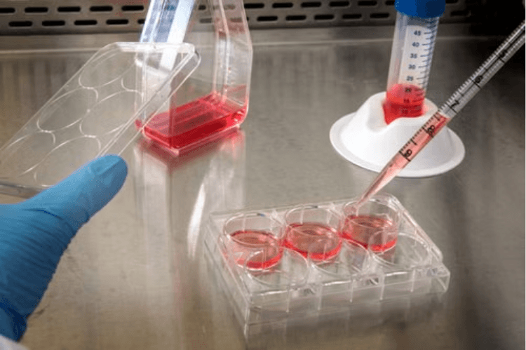
Biological contaminants include living organisms like bacteria, fungi, and viruses. They’re the most troublesome infectious agents, due to their ability to replicate fast.
Chemical contaminants encompass non-living agents. Examples include;
Bacteria are arguably the leading cause of tissue culture contamination. Common culprits include E.coli, Campylobacter, and Salmonella.
Mycoplasma is a sub-microscopic bacterial relative responsible for a significant percentage of tissue culture contamination.
This organism lacks a cell wall, making it resistant to standard antibiotics and difficult to detect under regular microscopes.
Molds account for the vast majority of fungal contamination cell culture agents.
Because these contaminants are significantly larger than bacteria, they’re much easier to detect.
Infectious viruses may invade tissue cultures and alter cell behaviors. Like mycoplasma, viral contaminants can replicate for weeks unnoticed.
Considered one of the most unique cell culture contamination types, cross-contamination occurs when rapidly proliferating cell lines aggressively attack other cells within a medium or incubator. HeLa cells are a notable culprit here.
Chemical tissue culture contamination often results from non-living impurities, such as heavy metals and detergent residues.
The toxic agents can inhibit cell growth and even trigger mass cell apoptosis.
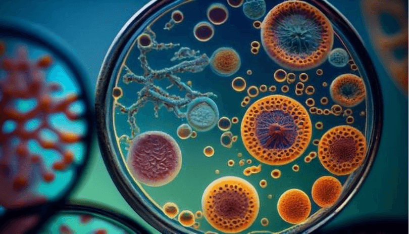
Always quarantine new tissue culture samples or cell lines before introducing them to the main laboratory environment.
Also, screen the cultures regularly for hidden contaminants. Conducting specialized tests like PCR can reveal slow-growing pathogens ahead of time, safeguarding your established stock.
Discard contaminated samples immediately and inform your supplier.
The use of antibiotics in tissue culture has garnered significant attention in the recent past, a nod to their efficacy.
Fungal invaders require a different approach. Products like Amphotericin B are commonly added to culture media to suppress fungal and yeast contamination, complementing standard antibacterial antibiotics.
Antibiotics are most effective against bacterial pathogens. They kill unwanted microbes, allowing the target cells and tissues to grow unimpeded.
However, only use antibiotics selectively and preferably during a cell culture’s initial stages. Prolonged exposure can lead to bacterial resistance.
Practicing strict aseptic protocols can minimize tissue culture contamination by creating a physical barrier between cell media and the external, microbe-rich environment. Some best practices you can implement here include;
While this may seem like a no-brainer, it’s one area where many researchers skimp. And the consequences can be devastating.
As a general practice, disinfect your workstation before and after every session. That goes for countertops, PPE, culture vessels, and other items entering the workspace.
Some effective sanitization techniques include;
Using properly sealed, sterile labware — such as centrifuge tubes rated for aseptic handling further reduces the risk of introducing contaminants during sample transfer and storage.
Always conduct your experiments inside the hood. The hood provides a stable unidirectional airflow, preventing microbes from flowing back and settling onto sensitive media samples.
Remember to also keep the laminar flow hood clean. Especially maintain the air vents to avoid harmful backflow of potential toxins.
To further prevent contamination in tissue culture, run the hood’s ultraviolet (UV) sterilization lamp after each session. It helps disinfect surfaces and items.
As a bonus point, only handle one tissue sample and focus on a single workflow at a time. This minimizes cross-contamination, especially while dealing with aggressive cell lines like HeLa.
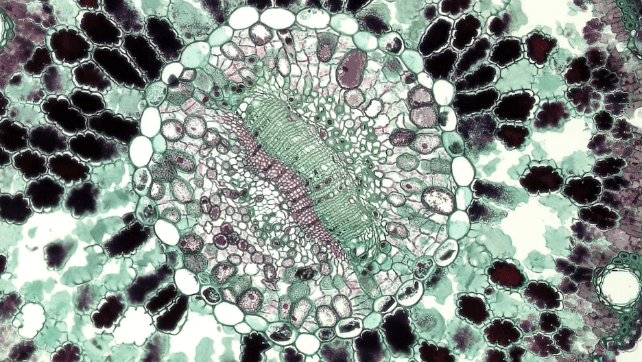
The consequences of tissue culture contamination are too grim to contemplate. Often, a single invader is all it takes to derail or ruin an important experiment.
The good news is - tissue culture contamination is completely preventable.
Maintaining a clean and sterile workspace can help stop invaders in their tracks. Besides, experts recommend isolating new cell lines for evidence of pre-contamination.
XL Biotec supplies essential reagents designed to prevent tissue culture contamination, including antifungal agents, sterile labware, and detection tools to screen for early signs of infection. Explore our full range of tissue culture solutions and find a product suited to your next experiment.
Explore our extensive collection of tissue culture solutions and discover a product best suited for your next experiment.
Bacteria, mycoplasma, and fungi are the main types of tissue culture contamination. Contamination could also result from viruses or chemical invaders, such as heavy metals.
Common physical signs of tissue culture contamination include cloudiness or discoloration.
Depending on the invader, you may also notice surface films or cottony masses in your cell nutrient medium.
Different types of bacteria can contaminate your tissue culture, ranging from E.coli to Campylobacter.
Mycoplasma doesn’t cause immediate media discoloration or dramatic pH shifts, making it potentially difficult to detect.
Effective detection requires using specialized enzyme assays or PCS testing.
Antibiotics can prevent tissue culture contamination by extending the longevity of the cell medium. They’re most effective against bacterial infections.
Cross-contamination, within the context of tissue cultures, occurs when an aggressive cell line overshadows other cells. This may happen within the same media, incubator, or workspace.
Practicing strict aseptic techniques can prevent tissue culture contamination by creating an impenetrable barrier between the nutrient medium and the external, microbe-rich environment.
Whether you can rescue a contaminated culture depends on its type. Plant-based tissues are generally salvageable, while contaminated mammalian cells are often difficult to redeem.
More importantly, success depends on how early you detect the invasion.
Conducting weekly screening helps detect mycoplasma early enough.
Remember, this bacterial relative can fly under the radar for weeks and cause significant contamination unnoticed.
The laminar-flow hood prevents tissue culture contamination by creating a physical barrier against external microbes. It ensures a stable unidirectional airflow, preventing airborne contaminants from flowing back into your cell nutrient medium.
