Your list is empty
Add products to request a quote

Somewhere between 15 and 35 percent of cell culture labs are running a mycoplasma-positive culture at any given time, and most don't know it. The cells look normal under the microscope, but the data quietly drifts anyway.
That single statistic explains why RPMI 1640 troubleshooting has to go beyond checking whether the medium looks pink or yellow that morning.
RPMI 1640 is one of the most popular cell culture media for lymphocytes, hybridomas, and suspension cell lines. It’s also one of the most unforgiving when something in your process goes wrong—glutamine spontaneously degrades, pH changes quickly in a crowded flask, and supplements that stimulate one cell line don’t support growth in another line.
Let’s walk you through the most common failure points we see researchers hit and how to fix them before they cost you a passage, plate, or dataset.
RPMI 1640 troubleshooting is a method for determining why a culture is not performing as expected and correcting the specific variable at fault, rather than discarding the entire medium and hoping the next lot will be better.
Because it’s a fully defined base medium, most problems can be traced back to a small set of variables: pH drift, glutamine breakdown, contamination, storage age, or a supplement mismatch for your specific cell type.
Researchers working with RPMI 1640 for tissue culture generally find that isolating one variable at a time, rather than changing the entire protocol, resolves the issue more quickly and keeps the rest of the culture history usable for comparison.
Cell growth can be affected by pH crashes, glutamine degradation, microbial contamination, and nutritional amendments. Here is an overview of each symptom, likely cause, and what to check first when performing RPMI 1640 troubleshooting:
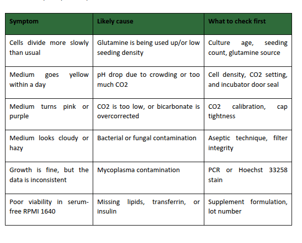
RPMI 1640 pH issues medium relies on a sodium bicarbonate buffer. This chemical system maintains a pH between 7.2 and 7.4. It needs a 5% CO₂ incubator to work.
So, move that flask out of the incubator too often, leave the cap too loose, or let cell density climb past what the flask can support, and the phenol red indicator turns yellow within hours.
A shift toward pink or purple usually means the same thing as above, but in reverse: either CO2 is too low, or the cap is not adequately screwed on to allow for proper gas exchange.
Adjusting in either direction affects enzyme and membrane transport activities, and cultures left out of range for more than a few hours will generally have reduced viability by the next passage, even if they look good that day.
Glutamine is the major energy source for rapidly dividing cells in this medium, but it is chemically unstable in solution.
It degrades unexpectedly into ammonia and pyroglutamate, and the breakdown speeds up with every freeze-thaw cycle and with every week the complete medium sits at 4 degrees Celsius.
This is the single biggest reason why a culture that grew well for months suddenly slows down without any apparent reason. The media looks clear, the pH looks fine, but the cells aren't dividing as fast as they were, and it's because of the accumulation of ammonia byproducts.
Switching to a stabilized form such as GlutaMAX or adding fresh L-glutamine weekly instead of relying on a large pre-made batch removes the variable entirely.
Mycoplasma cells measure between 0.15 and 0.3 micrometers, small enough to pass through a standard 0.22-microliter filter. That means contamination can enter through the medium itself, not just through a break in aseptic technique.
A mycoplasma contamination review published in PMC found that infection alters cell metabolism and growth rate without causing the turbidity or color change associated with bacterial or fungal contamination, which is exactly why it goes undetected for so long.
If growth has slowed and there’s nothing you can link it to, treat mycoplasma contamination as the default suspect and confirm with a PCR-based detection kit or Hoechst 33258 staining rather than relying on visual inspection alone.
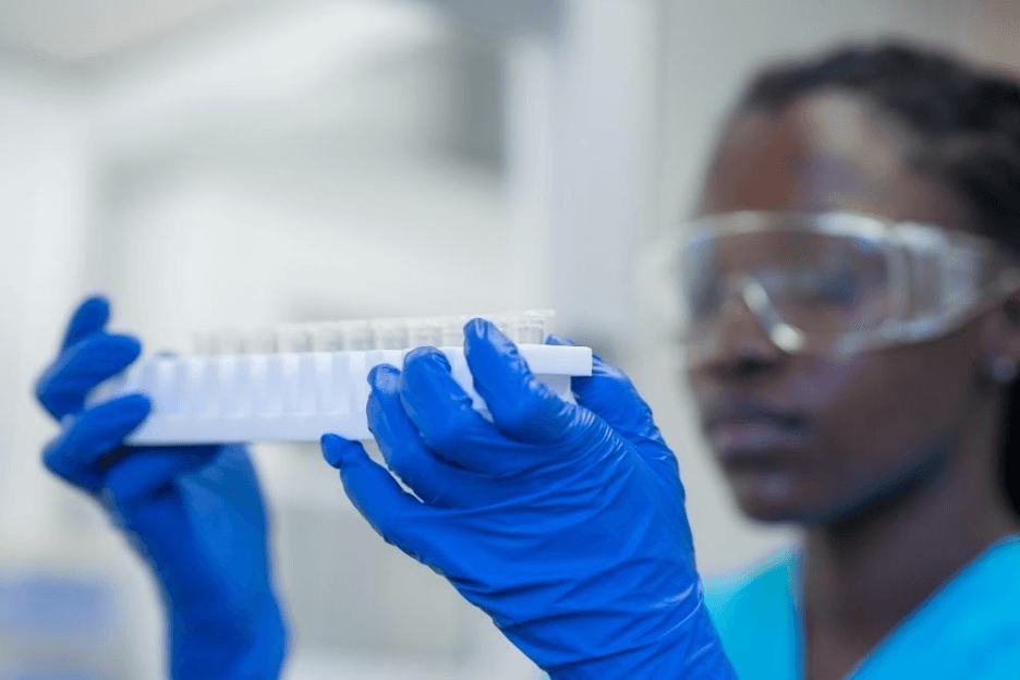
This is the foundation of much of immunology and oncology, from PBMC assays to cell line maintenance for Jurkat, K-562, and HL-60. One wrong supplement or missed contamination check doesn’t contaminate a single flask, but many potentially.
It can render inter-passage comparisons void, distort drug screening results, or compel a lab to dump a hybridoma that’s taken several months to generate.
Reproducibility depends on catching these problems early. Cytokine-dependent assays, in particular, are so sensitive to even slight drifts in medium quality. That’s why many labs running T-cell or hybridoma work will further pair them with tested lots of cytokines and growth factors, rather than risk having to swap suppliers mid-protocol.
RPMI 1640 Supplements that Solve Growth Problems
Most supplement issues come down to three additions: 10 percent FBS, 2 mM L-glutamine or GlutaMAX, and 1 percent penicillin-streptomycin.
Beyond that baseline, sodium pyruvate and beta-mercaptoethanol are worth adding for cell types that are sensitive to oxidative stress, including many primary lymphocyte cultures.
A serum-free version removes FBS, improving lot-to-lot consistency and reducing the risk of contamination from animal-derived components. However, FBS also provides lipids, transferrin, and insulin, so a serum-free formulation must directly complement those components, or growth will stall, no matter how technically correct the base medium.
Labs that begin moving to serum-free, especially if it's for cell therapy or GMP-adjacent work, will have an adaptation period. It can often take 2-3 passages for cells used to serum to return to the same growth rate.

T cell culture adds a few specific requirements on top of the standard base: Beta-mercaptoethanol at around 50 micromolar supports proliferation, particularly under serum-free or low-serum conditions, by maintaining the intracellular redox balance T cells need to divide.
IL-2 is typically added at concentrations of 10-600 IU/mL, depending on the protocol. However, higher doses can reduce cytotoxicity even as they improve proliferation, so the concentration is better tuned than maximized.
Antibody-based activation (usually anti-CD3 and anti-CD28) is added to the supplemented medium to drive expansion. Getting the base medium right before adding activation reagents makes it much easier to tell whether a poor expansion result is a stimulation problem or a medium problem.
Most RPMI 1640 problems trace back to a handful of variables: pH stability, glutamine freshness, contamination, and a supplement plan matched to the cell type in the flask. Working through them in that order, rather than replacing the entire protocol at the first sign of trouble, saves time and difficult-to-replace cell lines.
XL Biotec supplies RPMI 1640 and a full range of tissue culture products for labs across Thailand.
If you're setting up a new protocol or replacing a batch that has been giving you trouble, request a quote, and our team can help you match the right formulation and supplements to your cell line.
1. Why are my cells not growing in RPMI 1640 even though the medium looks normal?
A seemingly normal medium can still easily have bad glutamine, a pH that drifted below 7.2 or above 7.4 between tests, or a mycoplasma infection, none of which would cause the medium to be discolored. First, rule out the age of the glutamine and pH, as both are easy to test for quickly, and then move on to mycoplasma PCR testing if you still can’t get any growth.
2. How do I know if my RPMI 1640 has gone bad?
Check the expiration date and storage temperature first; a complete formulation containing glutamine should be used within 4 to 6 weeks of preparation and stored at 4 degrees Celsius, away from light. A color shift toward deep yellow or purple, visible particulate, or a sudden change in growth rate across multiple flasks at once are all signs that the batch should be discarded rather than further troubleshot.
3. Can I fix pH problems in RPMI 1640 without killing my entire batch?
Yes, in most cases. If the media has only drifted slightly and shows no other evidence of contamination, re-equilibrating in the CO2 incubator with the cap loose for 30-60 minutes will often bring it back into target range. A larger drift is usually an indication that you have too many cells or a problem with the CO2 calibration, and not something that the media can fix.
4. Can I fix pH issues in RPMI 1640 without remaking the whole batch?
Yes, in most cases. If the medium has only drifted slightly and shows no sign of contamination, re-equilibrating in the CO2 incubator with the cap loosened for 30 to 60 minutes often restores the target range. A more significant drift is usually a sign of overcrowding or a CO2 calibration issue rather than something the medium itself can fix.
5. How often should I test for mycoplasma contamination in RPMI 1640?
Monthly testing is generally recommended for active cultures, and any cell line entering the lab should be tested before being introduced into shared incubator space, as mycoplasma contamination will quickly spread between cultures once it gains access.
